


Renal abscess: A sinus tract extending to the skin surface
Submitted by: Mr Stephen Moore (Sonographer), Greater Glasgow and Clyde NHS Scotland. Contributing Author: Miss Heather MacDonald (Trainee Sonographer)
Patient Presentation/ Clinical information:
A 65 year old male presented for renal ultrasound after presenting with a large palpable mass located on the posterior left flank which had grown in size over the last week. The patient’s biochemistry was grossly normal however his CRP was elevated. He had a previous medical history of chronic left sided hydronephrosis and chronic liver disease, however no further history of note.
B-mode ultrasound demonstrated gross left sided hydronephrosis with thinning of the renal cortex which is in keeping with previous imaging (fig1). However, deep to the clinically obvious swelling on the left flank there was an irregular heterogeneous fluid collection which extended from the superficial soft tissues/skin to the lower pole region of the left kidney with which it appeared intimately associated (fig2, fig3 and Fig 4). These findings were concerning for a large renal abscess which may have progressed to a perirenal abscess and developed a sinus tract extending to the skin surface. In light of the elevated CRP, urgent cross-sectional imaging and urgent clinical review by the urology team was conducted. The right kidney and urinary bladder were unremarkable on ultrasound.
Computed Tomography (CT) confirmed the ultrasound findings of a large, multiloculated collection extending posteriorly from the left kidney through several fascial planes to the skin surface (Fig5). CT report stated that while this may be suggestive of an extensive renal abscess development with a sinus tract extending to the skin surface, a malignancy remained a differential.
 |
| Figure 1: Left sided hydronephrosis with renal cortical thinning (Star: Dilated collecting system, Triangle: Thinned cortex) |
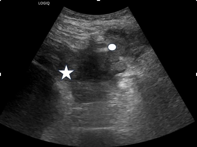 |
| Figure 2: Lower pole of the left kidney with the abscess seen extending into the extrarenal space (Star: Lower pole of the left kidney, Circle: Sinus tract) |
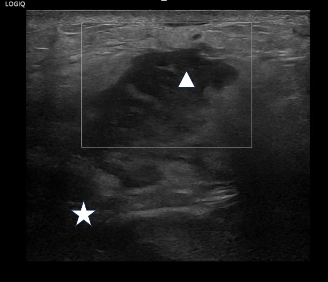 |
| Figure 3: Sinus tract extending from the left kidney through the posterior abdominal fascial planes (Star: Intraperitoneal aspect of the sinus tract, Triangle: Subcutaneous aspect of the sinus tract) |
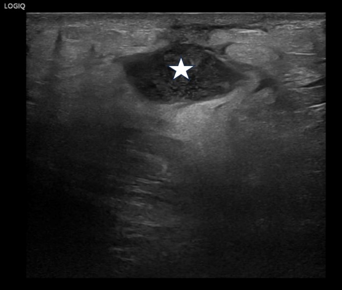 |
| Figure 4: High resolution linear probe image of the palpable lump demonstrating pyogenic content with low level echogenic debris (Star: Collection with in the subcutaneous tissues) |
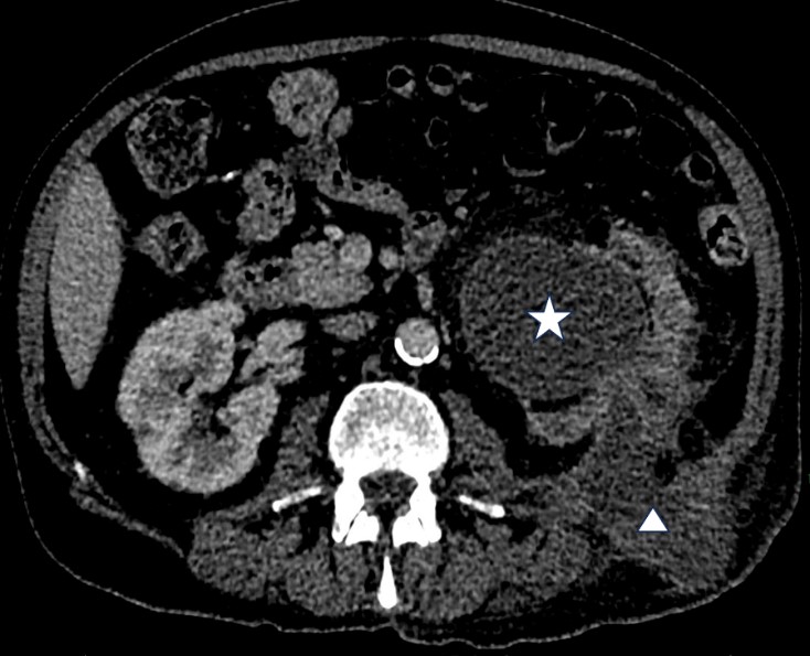 |
| Figure 5: CT image of the left renal abscess extending through the posterior abdominal wall facia to the dermis of the left flank. (Star: Hydronephrotic kidney, Triangle: Sinus tract extending through the posterior musculature of the abdominal wall to the skin surface) |